
What is mechanical ventilation?
Mechanical ventilation is a life-support therapy that uses an automated machine called a ventilator to assist or fully replace breathing when a person's respiratory system cannot maintain adequate oxygenation or carbon dioxide removal on its own. The ventilator acts as bellows, pushing warm, moist air or oxygen-enriched air into the lungs using positive pressure, then allowing the lungs to passively exhale. It does not treat the underlying illness. Instead, it stabilizes the patient while medications and other treatments address the root cause.
Mechanical ventilation is not the same as intubation. Intubation is the act of placing a breathing tube into the airway. Ventilation is what happens after that tube is connected to the machine. You can think of intubation as the access point and mechanical ventilation as the ongoing therapy delivered through it.
Key facts about mechanical ventilation:
- A ventilator controls breath frequency, pressure, and volume, all tailored to the individual patient's needs
- About 20–30% of ICU patients require mechanical ventilation at any given time in the United States
- Ventilators are used in hospital ICUs, operating rooms, emergency transport vehicles, and occasionally in the home for long-term conditions
- The machine can either fully breathe for the patient or simply assist breaths the patient initiates
- Ventilation keeps airways open, delivers oxygen, and prevents lung segments from collapsing due to insufficient pressure
- It is a temporary bridge to recovery, not a permanent solution
What are the different types of mechanical ventilation?
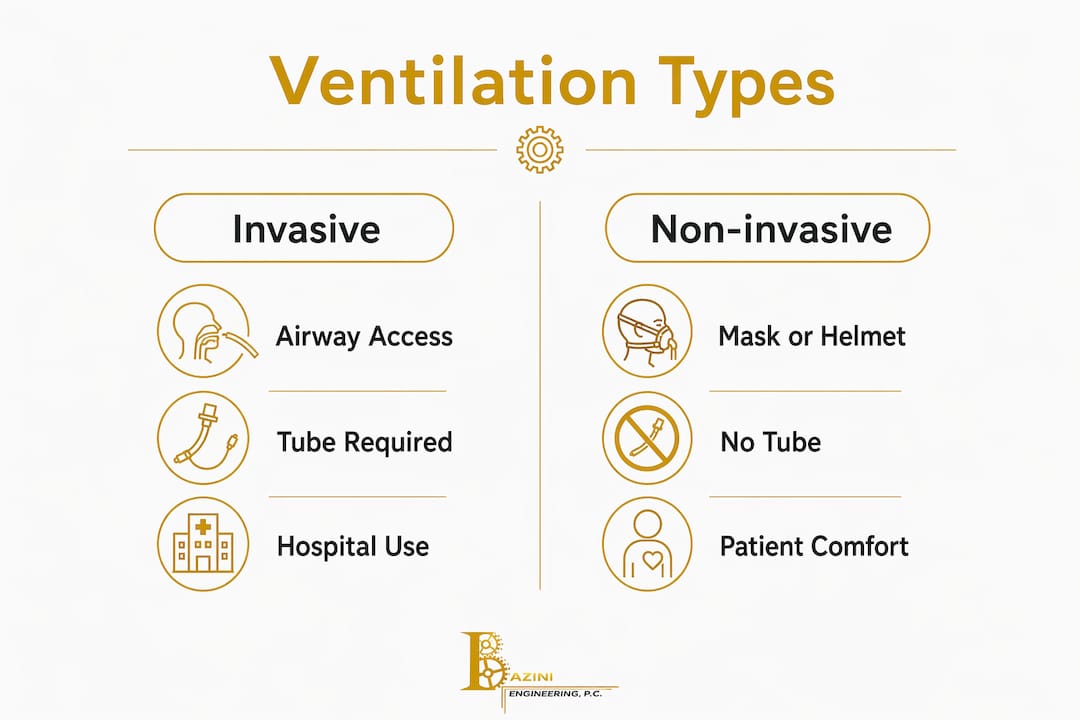
Mechanical ventilation falls into two broad categories: invasive and non-invasive. The choice between them depends on the severity of respiratory failure, the patient's level of consciousness, and how well the airway can be protected.
Invasive mechanical ventilation
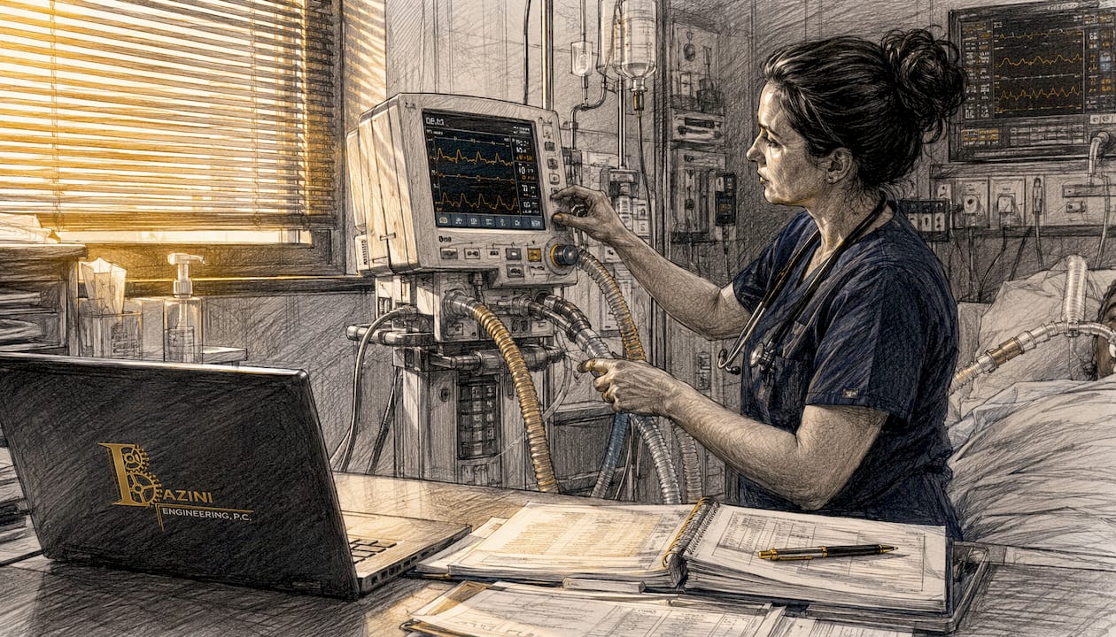
Invasive ventilation delivers positive pressure breaths through a tube placed directly into the airway. An endotracheal tube passes through the mouth or nose into the trachea and is secured at the lips. For patients who need ventilation longer than a few weeks, a tracheostomy tube is placed surgically through the front of the neck, which is more comfortable and easier to manage over time.
Non-invasive mechanical ventilation
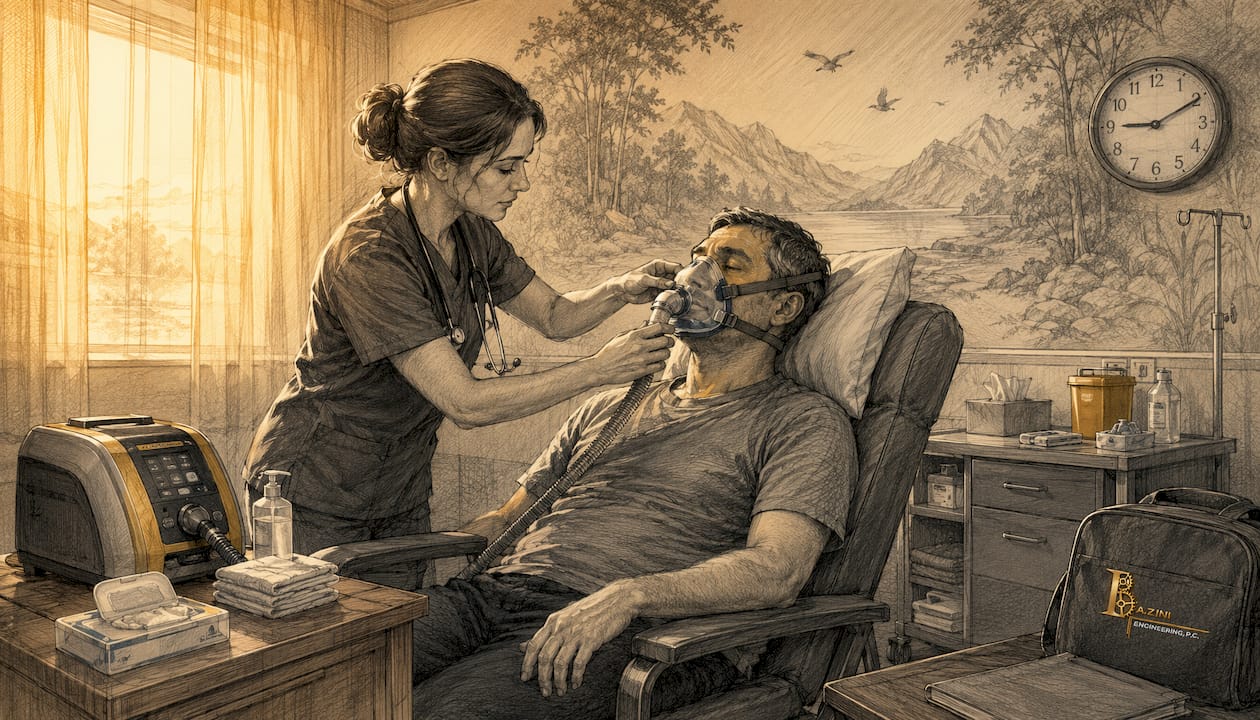
Non-invasive ventilation delivers positive airway pressure through a tightly fitting mask or helmet, with no tube entering the airway. Common forms include CPAP (continuous positive airway pressure) and BiPAP (bilevel positive airway pressure). CPAP delivers one constant pressure level, while BiPAP delivers a higher pressure during inhalation and a lower one during exhalation. These methods work well for patients who are conscious, cooperative, and able to protect their own airway.
Ventilation modes
Ventilators operate in several modes, each controlling how breaths are triggered and delivered:
- Assist-control (AC): The ventilator delivers a full breath every time the patient triggers it, and also delivers mandatory breaths if the patient's rate falls below the set minimum. This is the most common mode for critically ill patients.
- Pressure support ventilation (PSV): The patient triggers every breath, and the ventilator provides a set pressure boost to assist the effort. Used frequently during weaning.
- Pressure control ventilation (PCV): The ventilator delivers breaths at a fixed pressure, with volume varying based on lung compliance. Useful when limiting peak airway pressure is a priority.
- Synchronized intermittent mandatory ventilation (SIMV): Mandatory breaths are delivered in sync with the patient's own efforts, with spontaneous breaths allowed between them.
| Feature | Invasive ventilation | Non-invasive ventilation |
|---|---|---|
| Airway access | Endotracheal or tracheostomy tube | Mask or helmet |
| Level of respiratory failure | Severe or complete | Mild to moderate |
| Patient cooperation needed | No | Yes |
| Infection risk | Higher (pneumonia risk) | Lower |
| Typical setting | ICU, operating room | ICU step-down, emergency |
| Duration | Days to weeks | Hours to days |
Why is mechanical ventilation used, and who needs it?
Mechanical ventilation becomes necessary when the respiratory system fails to do one or both of its core jobs: getting enough oxygen into the blood (oxygenation) and removing carbon dioxide (ventilation). These failures can happen suddenly or develop over hours.
The most common clinical reasons a care team initiates mechanical ventilation include:
- Hypoxemic respiratory failure: Oxygen levels in the blood drop dangerously low despite supplemental oxygen, as seen in pneumonia, ARDS (acute respiratory distress syndrome), or pulmonary edema
- Hypercapnic respiratory failure: Carbon dioxide builds up because the patient cannot breathe deeply or fast enough, common in COPD exacerbations or neuromuscular disease
- Airway compromise: Trauma, burns, anaphylaxis, or a depressed level of consciousness leaves the airway at risk of obstruction or aspiration
- Surgical anesthesia: General anesthesia suppresses the drive to breathe, so the ventilator takes over for the duration of the procedure
- Cardiac arrest or severe shock: The body's oxygen demand outpaces what spontaneous breathing can supply
- Neuromuscular conditions: Diseases like ALS, Guillain-Barré syndrome, or high spinal cord injuries paralyze the breathing muscles
Patient groups most frequently requiring ventilation include critically ill adults in the ICU, post-surgical patients recovering from major procedures, premature infants with underdeveloped lungs, and trauma patients with chest injuries. Timing matters enormously. Delayed intubation in a deteriorating patient increases the risk of cardiac arrest during the procedure and worsens outcomes.
Pro Tip: Clinical teams watch for a combination of signs rather than a single number: rising respiratory rate, falling oxygen saturation, use of accessory neck muscles, and altered mental status together signal that ventilation may be imminent.
How is mechanical ventilation administered?
Starting mechanical ventilation is a coordinated process involving the physician, respiratory therapist, and nursing team. Each step builds on the last, and getting the initial settings right reduces complications downstream.
-
Secure the airway. For invasive ventilation, the physician performs rapid sequence intubation: sedation and a paralytic agent are given, the vocal cords are visualized with a laryngoscope, and the endotracheal tube is passed and confirmed with a chest X-ray and end-tidal CO2 monitoring. For non-invasive ventilation, the respiratory therapist fits the appropriate mask and adjusts the seal.
-
Set initial ventilator parameters. The respiratory therapist programs the machine based on the patient's height, weight, and diagnosis. Tidal volume, respiratory rate, fraction of inspired oxygen (FiO2), and PEEP are the four core settings adjusted at the start.
-
Confirm ventilation and oxygenation. Within 30 minutes of starting, an arterial blood gas (ABG) is drawn to check oxygen and carbon dioxide levels. The team adjusts settings based on results.
-
Monitor continuously. Nurses and respiratory therapists track peak airway pressure, plateau pressure, tidal volumes, and the patient's synchrony with the machine throughout each shift.
-
Perform airway suctioning. Secretions accumulate in the endotracheal tube and airways. Regular suctioning clears the tube and prevents mucus plugging, which can cause sudden drops in oxygen saturation.
-
Adjust settings as the patient's condition changes. Lung compliance and resistance shift with disease progression or recovery, so ventilator settings are continuously tailored by the healthcare team.
Key clinical protocols during ventilation:
- Elevate the head of the bed to 30–45 degrees to reduce aspiration risk
- Perform daily sedation interruptions to assess neurological status
- Use lung-protective ventilation strategies, particularly in ARDS patients
- Monitor cuff pressure on the endotracheal tube to prevent tracheal injury
- Coordinate oral care to reduce bacterial colonization in the mouth
How long does mechanical ventilation last, and what does weaning involve?
Duration varies widely. A patient ventilated during a routine surgery may be off the machine within hours of waking up. Someone with severe ARDS or a major neurological injury may need ventilation for weeks. There is no universal timeline because the underlying condition drives everything.
Weaning is the physiological process of gradually transferring the work of breathing from the ventilator back to the patient. It is not an abrupt event. The team reduces ventilator support in stages, watching closely for signs that the respiratory muscles are strong enough to sustain independent breathing.
Criteria clinicians use to assess readiness for weaning:
- The underlying cause of respiratory failure is improving or resolved
- The patient can initiate spontaneous breaths
- Oxygen requirements are low enough that FiO2 is at or below 0.4–0.5 with PEEP at or below 5–8 cm H2O
- The patient is awake enough to follow simple commands
- Hemodynamic stability without high-dose vasopressor support
- Secretion burden is manageable without frequent deep suctioning
Once the patient passes a spontaneous breathing trial (SBT), the team removes the breathing tube in a process called extubation. Sedation management is central to weaning. Excessive sedation suppresses the respiratory drive and delays the process, while inadequate sedation causes agitation, increased oxygen consumption, and patient-ventilator dyssynchrony. Weaning also requires attention to psychological comfort, since anxiety itself increases the body's oxygen demand and can stall progress.
What are the risks and complications of mechanical ventilation?
Mechanical ventilation is a life-saving intervention, but it carries real risks. The longer a patient remains on the ventilator, the higher the cumulative exposure to those risks. Understanding them helps caregivers ask the right questions and helps clinical teams prioritize prevention.
Common complications include:
- Ventilator-associated pneumonia (VAP): Bacteria colonize the endotracheal tube and migrate into the lungs. VAP is one of the most serious ICU infections and prolongs hospital stays.
- Barotrauma: Excessive airway pressure can rupture alveoli, causing air to leak into the pleural space (pneumothorax) or surrounding tissues.
- Oxygen toxicity: Prolonged exposure to high concentrations of supplemental oxygen damages lung tissue at the cellular level.
- Vocal cord damage: The endotracheal tube passes between the vocal cords, and prolonged intubation can cause hoarseness, granulomas, or, rarely, permanent scarring.
- Diaphragm weakness: The diaphragm atrophies quickly when the ventilator does all the work, making weaning harder the longer ventilation continues.
- Psychological effects: Patients who are awake during ventilation often experience fear, disorientation, and post-traumatic stress after ICU discharge.
Mechanical ventilation is a bridge, not a destination. The goal from day one is to reduce support as fast as the patient's physiology allows, because every additional day on the machine adds risk without adding benefit once the underlying condition is stabilizing.
Monitoring and early detection are the primary defenses against these complications. Lung-protective ventilation strategies, strict hand hygiene, oral care bundles, and daily sedation interruptions all reduce the incidence of the most serious problems. The clinical team's job is not just to keep the patient breathing but to get them off the ventilator as safely and quickly as possible.
How does mechanical ventilation work physiologically?
Natural breathing works on negative pressure. When the diaphragm contracts and the chest wall expands, pressure inside the lungs drops below atmospheric pressure, and air flows in passively. Exhalation is passive: the elastic recoil of the lungs pushes air out. Mechanical ventilation reverses this entirely.
A ventilator uses positive pressure to push air into the lungs, overcoming the resistance of the airways and the stiffness of the lung tissue. The two key mechanical properties that govern how the lungs respond are compliance (how easily the lungs stretch) and resistance (how much the airways oppose airflow). Stiff lungs, as in ARDS, have low compliance and require higher pressures to achieve adequate tidal volumes. Obstructed airways, as in asthma or COPD, have high resistance and require longer expiratory times to empty fully.
The four phases of a ventilator breath
Each breath cycle has four distinct phases, and clinicians adjust settings within each:
- Trigger phase: The breath begins, either because the patient makes an inspiratory effort (patient-triggered) or because the set time interval has elapsed (time-triggered). Trigger sensitivity is adjusted to match the patient's effort without causing the machine to fire on its own.
- Inspiratory phase: The ventilator delivers gas at the set flow rate, pressure, or volume. This is where tidal volume and peak pressure are determined.
- Cycling phase: The ventilator switches from inhalation to exhalation. Cycling can be triggered by time, volume, flow rate, or pressure, depending on the mode.
- Expiratory phase: The patient exhales passively. The ventilator does not actively pull air out; it simply opens the expiratory valve and allows elastic recoil to do the work.
PEEP and pressure management
Positive end-expiratory pressure (PEEP) is the pressure maintained in the airways at the end of exhalation, preventing alveoli from collapsing between breaths. In conditions like ARDS, where alveoli are prone to collapse, therapeutic PEEP dramatically improves oxygenation by keeping more lung tissue open and participating in gas exchange.
A separate phenomenon called intrinsic PEEP, or auto-PEEP, occurs when the lungs do not fully empty before the next breath begins. Auto-PEEP builds up alveolar pressure unintentionally and increases the work the patient must do to trigger the ventilator, which can fatigue the respiratory muscles and complicate weaning. Distinguishing between applied PEEP and auto-PEEP is a routine part of ventilator management in any ICU.
Pressure management is where mechanical ventilation gets genuinely complex. A setting that improves oxygenation in one patient can cause barotrauma in another. The difference comes down to individual lung mechanics, which is why continuous monitoring and real-time adjustment are non-negotiable in critical care.
| Physiological concept | What it means clinically |
|---|---|
| Lung compliance | How easily the lung stretches; low compliance requires higher pressure to deliver the same volume |
| Airway resistance | Opposition to airflow; high resistance requires longer expiratory time to prevent air trapping |
| Therapeutic PEEP | Applied pressure at end-exhalation to keep alveoli open and improve oxygenation |
| Auto-PEEP | Unintended pressure buildup from incomplete exhalation; increases patient effort and risk |
| Tidal volume | Volume of air per breath; set relative to ideal body weight to protect lung tissue |
Understanding ventilation pressure principles helps caregivers grasp why the clinical team makes specific adjustments and why those decisions directly affect how quickly a patient recovers.
Key Takeaways
Mechanical ventilation is a positive-pressure life-support therapy that stabilizes breathing, protects oxygenation, and buys time for recovery while the underlying condition is treated.
| Point | Details |
|---|---|
| Definition and purpose | A ventilator uses positive pressure to move air into the lungs when spontaneous breathing fails or is unsafe. |
| Two main types | Invasive ventilation uses an endotracheal or tracheostomy tube; non-invasive uses a mask or helmet. |
| ICU prevalence | About 20–30% of ICU patients in the United States require mechanical ventilation at any given time. |
| Weaning is gradual | Discontinuing ventilation is a staged process requiring respiratory recovery, sedation management, and spontaneous breathing trials. |
| Risks require active management | Ventilator-associated pneumonia, barotrauma, and diaphragm weakness are real risks managed through lung-protective strategies and daily reassessment. |
Mechanical engineering expertise you can trust

Ventilation principles extend well beyond the ICU. Whether you are designing HVAC systems for a hospital, a commercial building, or a residential complex, the underlying physics of airflow, pressure, and air quality follow the same rules. Baziniengineering brings that depth of mechanical engineering knowledge to every project it takes on.
As a licensed MEP engineering firm serving New York City, Long Island, Westchester County, and surrounding regions, Baziniengineering designs HVAC systems, plumbing networks, and fire protection systems that meet current code requirements and perform reliably in real-world conditions. The firm's mechanical engineering services cover everything from initial design through permit coordination with the NYC Department of Buildings and FDNY.
If your project requires code-compliant mechanical systems built on sound engineering principles, explore Baziniengineering's full services to see how the firm can support your next project.